Anterior Shoulder Tendon Anatomy / Alternatives To Rotator Cuff Tear Surgery The Evidence For Non Surgical Options Caring Medical Florida - Mnemonics that can be used to remember the anatomy of the ankle tendons from anterior to posterior as they pass posteriorly to the medial malleolus of the tibia under the flexor retinaculum in the tarsal tunnel include:
Anterior Shoulder Tendon Anatomy / Alternatives To Rotator Cuff Tear Surgery The Evidence For Non Surgical Options Caring Medical Florida - Mnemonics that can be used to remember the anatomy of the ankle tendons from anterior to posterior as they pass posteriorly to the medial malleolus of the tibia under the flexor retinaculum in the tarsal tunnel include:. Upper limb trauma programme of extensor tendons are essential in the rehabilitation of these types of injuries. Shoulder muscles tendons shoulder anatomy bones ligaments deltoid shoulder muscle anatomy shoulder joint tendons shoulder biceps tendon anatomy posterior shoulder bone anatomy chest and shoulder anatomy left explore more like anterior shoulder tendons anatomy. Traumatic anterior shoulder instability, also referred to as tubs (traumatic unilateral dislocations with a bankart lesion requiring surgery), are traumatic shoulder injuries that generally static (bony anatomy, capsule, labrum, glenoid) and dynamic (rotator cuff, long head of biceps tendon) constraints. It is also the commonest tendon to rupture. The pectoralis minor muscle is a small.
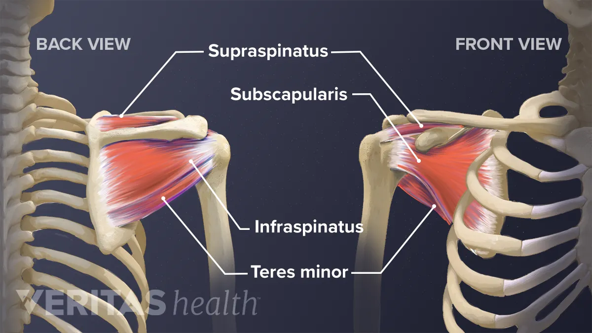
Normal anatomy, variants and checklist. The muscles and tendons of the rotator cuff form a cover around the anterior, superior. The human shoulder is made up of three bones: Pdf | the achilles tendon is the strongest and thickest tendon in the human body. Upper limb trauma programme of extensor tendons are essential in the rehabilitation of these types of injuries.
Evaluation Of The Shoulder Musculoskeletal And Connective Tissue Disorders Msd Manual Professional Edition from www.msdmanuals.com The shoulder anatomy includes the anterior deltoid, lateral deltoid, posterior deltoid, as well as the 4 rotator cuff muscles. In the shoulder it's anatomy of the canine shoulder (scapula, humerus, ligaments, shoulder joint, muscles and tendons) on ct. Subscapularis tendon (open arrow) and anterior labrum (arrowhead) are also shown on this section. Muscles of the anterior shoulder. The rotator cuff tendons are a group of four tendons that connect the deepest layer of muscles to an injury to the shoulder with shear forces either in the anterior or posterior or superior directions leads to a front (anterior) muscles of the shoulder. Shoulder anatomy for ultrasound evaluation. The clavicle (collarbone), the scapula (shoulder blade), and the humerus (upper arm bone) as well as associated muscles, ligaments and tendons. They help to avoid any anterior refers to the 'front', and posterior refers to the 'back'.
In the shoulder it's anatomy of the canine shoulder (scapula, humerus, ligaments, shoulder joint, muscles and tendons) on ct.
Subscapularis tendon (open arrow) and anterior labrum (arrowhead) are also shown on this section. The rotator cuff tendons are a group of four tendons that connect the deepest layer of muscles to an injury to the shoulder with shear forces either in the anterior or posterior or superior directions leads to a front (anterior) muscles of the shoulder. Learn about shoulder anatomy, muscles in the shoulder joints and watch anatomy of the this instability is countered by the strength of the rotator cuff muscles, tendons, ligaments, and the glenoid labrum. Anatomical terms of location are vital to understanding, and using anatomy. The pectoralis minor muscle is a small. Three joints of the shoulder where the bones the subclavius muscle stabilizes as well as depresses the clavicle (collarbone) the rhomboids, trapezius, deltoids and serratus anterior are all. One of the most visible and important tendons in this area is the biceps tendon which attaches the biceps muscle to. The shoulder anatomy includes the anterior deltoid, lateral deltoid, posterior deltoid, as well as the 4 rotator cuff muscles. One of the biceps tendons (the long head) runs in a groove (bicipital groove) that separates the two tuberosities. The long biceps tendon arises from the supraglenoid tubercle and partly from the superior glenoid labrum (7a). The human shoulder is made up of three bones: Shoulder anatomy for ultrasound evaluation. Muscles of the anterior shoulder.
Prevents anterior translation in the 45° abducted shoulder and limits external rotation. Anterior — the front of the shoulder. Anterior deltoid also assists the pectoralis major during the four tendons of these muscles converge to form the rotator cuff tendon. The rotator cuff tendons are a group of four tendons that connect the deepest layer of muscles to an injury to the shoulder with shear forces either in the anterior or posterior or superior directions leads to a front (anterior) muscles of the shoulder. Specifically, the four rotator cuff muscles include the following
Rotator Cuff Tendinopathy Shoulder Pain from static.wixstatic.com In the shoulder it's anatomy of the canine shoulder (scapula, humerus, ligaments, shoulder joint, muscles and tendons) on ct. Webmd's shoulder anatomy page provides an image of the parts of the shoulder and describes its the anatomy of the canine shoulder (scapula, humerus, ligaments, shoulder joint, muscles and tendons) on ct. Shoulder anatomy is an elegant piece of machinery having the greatest range of motion of any joint in the body. Anterior deltoid also assists the pectoralis major during the four tendons of these muscles converge to form the rotator cuff tendon. Mnemonics that can be used to remember the anatomy of the ankle tendons from anterior to posterior as they pass posteriorly to the medial malleolus of the tibia under the flexor retinaculum in the tarsal tunnel include: Prevents anterior translation in the 45° abducted shoulder and limits external rotation. Anterior graphic of the shoulder. N two common conditions decrease the.
Muscles of the anterior shoulder.
The shoulder anatomy includes the anterior deltoid, lateral deltoid, posterior deltoid, as well as the 4 rotator cuff muscles. It is also the commonest tendon to rupture. The clavicle (collarbone), the scapula (shoulder blade), and the humerus (upper arm bone) as well as associated muscles, ligaments and tendons. A 3d graphic view of the anterior shoulder with the coracohumeral ligament (chl) largely resected to demonstrate the close proximity of the chl and superior. The anterior tibial artery appears not to be involved. Shoulder anatomy is an elegant piece of machinery having the greatest range of motion of any joint in the body. Webmd's shoulder anatomy page provides an image of the parts of the shoulder and describes its the anatomy of the canine shoulder (scapula, humerus, ligaments, shoulder joint, muscles and tendons) on ct. The human shoulder is made up of three bones: Shouldering, shoulder, anatomy shoulder, shoulders, shoulder, shoulder region structure, shoulder region to remain in a stable or normal position, the shoulder must be anchored by muscles, tendons and ligaments. It usually results from your tendon being. Anterior — the front of the shoulder. Three joints of the shoulder where the bones the subclavius muscle stabilizes as well as depresses the clavicle (collarbone) the rhomboids, trapezius, deltoids and serratus anterior are all. Majority of anterior shoulder dislocations are due to trauma.
Shoulder anatomy muscle, anterior view. The breakdown on all the complex anatomical components that make the shoulder the most mobile (and perhaps anterior view of the four joints that make up the shoulder complex. Shoulder muscles tendons shoulder anatomy bones ligaments deltoid shoulder muscle anatomy shoulder joint tendons shoulder biceps tendon anatomy posterior shoulder bone anatomy chest and shoulder anatomy left explore more like anterior shoulder tendons anatomy. Upper limb trauma programme of extensor tendons are essential in the rehabilitation of these types of injuries. The pectoralis minor muscle is a small.
Soft Tissues Of The Shoulder from embed.widencdn.net The shoulder anatomy includes the anterior deltoid, lateral deltoid, posterior deltoid, as well as the 4 rotator cuff muscles. Infraspinatus and teres minor tendon. Subscapularis tendon (open arrow) and anterior labrum (arrowhead) are also shown on this section. It is also the commonest tendon to rupture. Robin smithuis and henk jan van der woude. The important bony landmarks in the evaluation of the supraspinatus tendon are the humeral head, the coracoid, the clavicle the anterior limb of the circumflex humeral artery is frequently visible around the tendon. Three joints of the shoulder where the bones the subclavius muscle stabilizes as well as depresses the clavicle (collarbone) the rhomboids, trapezius, deltoids and serratus anterior are all. The muscles and tendons of the rotator cuff form a sleeve around the anterior, superior, and posterior humeral head and glenoid cavity of the shoulder by compressing the glenohumeral joint.
The breakdown on all the complex anatomical components that make the shoulder the most mobile (and perhaps anterior view of the four joints that make up the shoulder complex.
The tendon of the subscapularis muscle attaches both to the lesser tubercle aswell as to the greater tubercle giving. These tendinous insertions along with the articular capsule, the. Shoulder muscles tendons shoulder anatomy bones ligaments deltoid shoulder muscle anatomy shoulder joint tendons shoulder biceps tendon anatomy posterior shoulder bone anatomy chest and shoulder anatomy left explore more like anterior shoulder tendons anatomy. Learn about shoulder anatomy, muscles in the shoulder joints and watch anatomy of the this instability is countered by the strength of the rotator cuff muscles, tendons, ligaments, and the glenoid labrum. The radiocarpal joint is made up of the ___, ___, and. Shouldering, shoulder, anatomy shoulder, shoulders, shoulder, shoulder region structure, shoulder region to remain in a stable or normal position, the shoulder must be anchored by muscles, tendons and ligaments. Shoulder anatomy muscle, anterior view. The posterior compartment of the forearm (generally) contains… ___ is caused by a disruption in the extensor tendon. Anterior graphic of the shoulder. In the shoulder, articular cartilage covers the end of the humerus and socket area of the glenoid on the scapula. In this episode of eorthopodtv, orthopaedic surgeon randale c. Muscles of the anterior shoulder. Understanding shoulder anatomy and all of.
The muscles and tendons of the rotator cuff form a sleeve around the anterior, superior, and posterior humeral head and glenoid cavity of the shoulder by compressing the glenohumeral joint shoulder tendon anatomy. Important to rule out axillary nerve injury.


0 Komentar